Bedside Brain Imaging: A New Era in Detecting Brain Injuries?
The brain is a wonderful thing. It allows us to process and understand the world around us, it allows us to communicate and to be able to move. The brain does things for us that we aren’t even conscious of, such as the regulating our breathing and heart rate. Our brain is responsible for almost everything we do, and as a result any damage to it can be devastating.
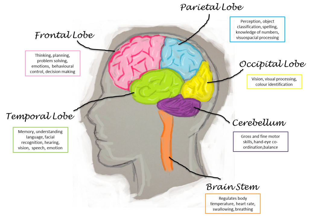
Around 1 million people present in A&E with some form of head injury in the UK, and although not all of these result in any long term problems there are around 500,000 people living in the UK with disabilities caused by their brain injury. The kind of disability that each person has depends on where their brain sustained an injury, as each lobe has a different function.
The quicker that injury to the brain is picked up the better the prognosis for the patient, and with this in mind scientists at Cambridge University have just been granted a sum of £800,000 to use towards research into bedside brain imaging techniques. The Cambridge Brain Injury Healthcare Technology Cooperative (HTC) are behind the project and hope to create a device that can take high quality images of a patient’s brain without them having to be put through a machine. They also hope to develop an app for the iPhone and iPad which is able to detect brain injury, which is something that will be welcomed within the medical world as this test with is currently done by doctors using pen and paper and takes up a lot of time.
What Causes Brain Injury?
There are many causes of brain injury, with some being more severe and having more lasting effects than others
- Acquired brain injury à this means that the injury was caused after birth and includes a stroke, choking, a virus that affects the central nervous system, heart attack, aneurysm, seizures, brain tumours and so on; the list is endless. These causes either involve a lack of oxygen to the brain, a disruption of the blood supply to the brain (which in turn results in a lack of oxygen,) or something that specifically acts on the brain to cause damage to it (a tumour or a virus.)
- Birth injury àBirth injury could be caused by a lack of oxygen during delivery, but many cases are due to medical negligence during the birth, which could be due to a number of things: failure to recognise foetal distress and plan for a C-section, traumatic use of surgical tools during delivery, or failing to recognise that a new born needs oxygen.
- Traumatic brain injury à this could include anything from a car crash or a fall to a sports injury or as a result of a violent fight.
The reason that quicker imaging of the brain is beneficial is that in some cases the amount of damage can be limited if treatment is started early. For example, if someone is having a stroke, a brain scan may show that an area of their brain has infarcted (died) due to a blockage of one of the arteries. In this case, a “clot-busting” drug called alteplase can be used to try and minimise the damage, but it is only effective for an hour and a half from the onset of the stroke.
Anatomy of the Brain
This diagram shows the locations of the different lobes of the brain and their individual functions.
What Symptoms Can We Expect if Each Lobe is Damaged?
So as you can see, the signs and symptoms that a patient presents with can alert you to the fact that they may have a brain injury, but by the time these signs show themselves it might be too late to repair the damage. A way of scanning the brain before these symptoms develop may save the patient and their family a lot of heartache and anguish, and give them the chance to live a normal life; a right that every human being has and quite rightly takes for granted.
[Guest Post by Niqui Stubbs, a 4th year medical student with a particular interest in medical negligence. She enjoys writing about this topic, and is currently working on behalf of a medical negligence specialist from the UK.]


